
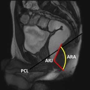
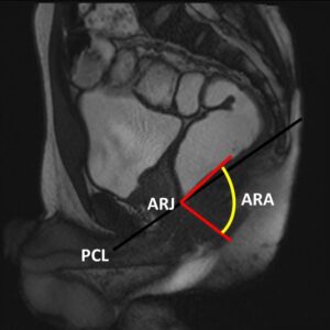
- Pubococcygeal line (PCL): connects inferior aspect of symphysis pubis to last coccygeal joint
- Anorectal juction (ARJ): crosspoint of line along posterior border of the rectum and line along central axis of anal canal
- Anorectal angle (ARA): Angle beween the two lines used to define the ARJ At rest base of bladder, upper third of vagina and peritoneal cavity should be above PCL. ARJ within 3cm below PCL.
- At squeeze elevation of pelvic organs and ARA should reduce by at least 15 to 20o
- At strain and defecation mild descent of pelvic organs (<2cm) expected. ARA typically increased by 15-20o more than rest
Measurements
 |
 |
Normal Study
| Rest | Squeeze |
|---|---|
| Strain | Defecation |
|---|---|
| Abnormality | Small | Moderate | Large |
|---|---|---|---|
| Rectal descent | < 3 cm* (normal) | 3-6 cm* | > 6 cm* |
| Bladder descent | < 3 cm* (normal up to 1 cm) | 3-6 cm* | > 6 cm* |
| Vagina descent | < 3 cm* | 3-6 cm* | > 6 cm* |
| Rectocele size | < 2 cm (normal) | 2-4 cm* | > 4 cm* |
| Enterocele | < 3 cm* | 3-6 cm* | > 6 cm* |
Abnormal Rectal Descent
|
|
Rectocele |
|
|
|
Rectal Intussusception |
|
|
|
Rectal Prolapse |
|
|
|
Anismus |
|
|
|
Enterocele |
|
|
|
Cystocele |
|
|
|
Utero-vaginal Prolapse |
|
 |
|
- Lalwani N, Moshiri M, Lee JH, Bhargava P, Dighe MK. Magnetic resonance imaging of pelvic floor dysfunction. Radiol. Clin. North Am. 2013 Nov;51(6):1127–39.
- Flusberg M, Sahni VA, Erturk SM, Mortele KJ. Dynamic MR defecography: assessment of the usefulness of the defecation phase. AJR Am. J. Roentgenol. 2011 Apr;196(4):W394–399.
- Mortele KJ, Fairhurst J. Dynamic MR defecography of the posterior compartment: Indications, techniques and MRI features. Eur. J. Radiol. 2007 Mar;61(3):462–72.
- Roos JE, Weishaupt D, Wildermuth S, Willmann JK, Marincek B, Hilfiker PR. Experience of 4 years with open MR defecography: pictorial review of anorectal anatomy and disease. Radiogr. Rev. Publ. Radiol. Soc. N. Am. Inc. 2002 Aug;22(4):817–3